- The study investigated cortical and autonomic nervous system activity during affective processing in post-traumatic stress disorder.
- Researchers used magnetoencephalography in 36 PTSD patients and 48 controls viewing positive and neutral images.
- PTSD was associated with attenuated early evoked response amplitude to all stimuli in right ventral and medial temporal regions.
- The authors concluded PTSD involves impaired early sensory processing, attentional control, and parasympathetic engagement.
- These identified alterations in cortical and autonomic activity may serve as targets for PTSD treatments.
Unpacking the Neural Signatures of Post-Traumatic Stress Disorder
Post-traumatic stress disorder (PTSD) is a prevalent and disabling condition, particularly among service members [1]. Its symptoms, including anhedonia and emotional numbing, significantly impair function and quality of life [2, 3, 4, 5]. While clinical management is guided by symptom presentation, the underlying neurobiology of these affective disturbances is not fully understood. A recent study used advanced neuroimaging to map the specific brain and autonomic nervous system dynamics that may contribute to these core symptoms, aiming to identify objective biological markers that could inform future diagnostics and treatments [6].
Investigating Affective Processing with Magnetoencephalography
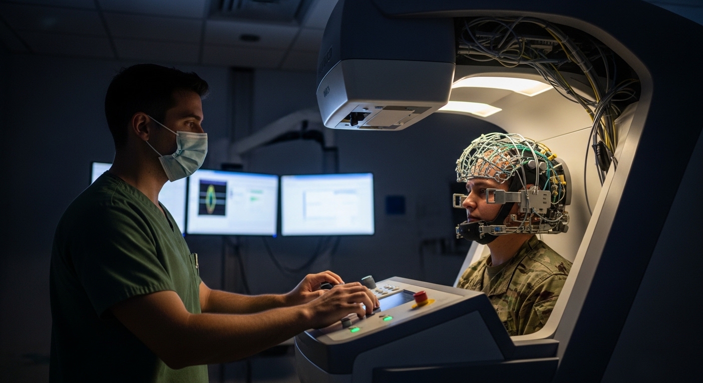
To detail the neural circuits of affective processing in PTSD, researchers utilized magnetoencephalography (MEG), a non-invasive technique that measures the minute magnetic fields produced by the brain's electrical activity. The high temporal resolution of MEG is particularly well-suited for tracking the rapid, millisecond-by-millisecond changes that characterize neural responses to emotional stimuli. The study cohort included 84 service members, who were assigned to one of two groups: 36 individuals who met DSM-V criteria for PTSD and 48 individuals who did not, serving as a control group.
Cortical Response Differences in PTSD
The investigation first established a baseline for healthy affective processing. In control participants, viewing pleasant images compared to neutral ones elicited higher amplitudes of early neuromagnetic evoked responses, occurring within 75 to 150 milliseconds post-stimulus. This rapid increase in activity across a distributed cortical network suggests a swift, automatic engagement with positively valenced information. In contrast, service members with PTSD demonstrated a markedly different pattern. They exhibited attenuated amplitude of these early evoked responses to all visual stimuli, including both positive and neutral images. This blunted neural response was most pronounced in the right ventral and medial temporal regions, areas of the brain critical for emotional processing and memory formation. This finding suggests an impairment in the initial, automatic stage of sensory processing in PTSD, potentially reflecting a diminished capacity of the appetitive motivational system to engage with environmental cues.
Altered Brain Oscillations and Autonomic Function
The study revealed further neural dysregulation in PTSD beyond the initial sensory response. During the task, individuals with PTSD showed less suppression of alpha and beta band power. In a healthy brain, the suppression of these brainwave frequencies indicates sustained attention and the filtering of irrelevant neural activity. The failure to adequately suppress these oscillations in the PTSD group points to an impairment in attentional control, which may manifest clinically as difficulty concentrating or being easily distracted. The investigation also extended to the autonomic nervous system, uncovering a distinct physiological signature. In response to all images, the PTSD group demonstrated delayed heart rhythm deceleration. This finding indicates a slower engagement of the parasympathetic nervous system, the body's primary system for calming and recovery. This sluggish autonomic response to environmental stimuli aligns with the broader pattern of physiological dysregulation characteristic of PTSD.
Clinical Implications: Connecting Findings to Symptoms and Treatment
The distinct patterns of neural and autonomic activity identified in this study provide a potential biological basis for the affective symptoms of PTSD. The impairment in early, automatic sensory processing, particularly in temporal lobe circuits, offers a neurophysiological explanation for the anhedonia and emotional numbing frequently reported by patients. This blunted initial response to the environment suggests that the brain in PTSD may not be registering the salience of potentially rewarding stimuli. This is compounded by the observed impairment in sustained attentional control, reflected by altered brain oscillations, which could make it difficult for individuals to remain engaged with their surroundings. Finally, the finding of a slower engagement of the parasympathetic system provides an objective measure of the autonomic dysregulation that contributes to a persistent state of hypervigilance and delayed emotional recovery. Together, these findings paint a cohesive picture of how disruptions in cortical and autonomic function may produce the clinical syndrome of PTSD. Crucially, these specific dysfunctions, from early evoked responses to heart rhythm modulation, can provide objective, quantifiable targets for treatments, allowing clinicians to potentially track the efficacy of interventions aimed at restoring sensory processing, attentional control, and parasympathetic tone.
References
1. Rogers J, Chesney E, Oliver D, et al. Psychiatric and neuropsychiatric presentations associated with severe coronavirus infections: a systematic review and meta-analysis with comparison to the COVID-19 pandemic. The Lancet Psychiatry. 2020. doi:10.1016/s2215-0366(20)30203-0
2. Salari N, Hosseinian‐Far A, Jalali R, et al. Prevalence of stress, anxiety, depression among the general population during the COVID-19 pandemic: a systematic review and meta-analysis. Globalization and Health. 2020. doi:10.1186/s12992-020-00589-w
3. Norman R, Byambaa M, De R, Butchart A, Scott JG, Vos T. The Long-Term Health Consequences of Child Physical Abuse, Emotional Abuse, and Neglect: A Systematic Review and Meta-Analysis. PLoS Medicine. 2012. doi:10.1371/journal.pmed.1001349
4. Wang J, Mann F, Lloyd‐Evans B, Ma R, Johnson S. Associations between loneliness and perceived social support and outcomes of mental health problems: a systematic review. BMC Psychiatry. 2018. doi:10.1186/s12888-018-1736-5
5. Kisely S, Warren N, McMahon L, Dalais C, Henry I, Siskind D. Occurrence, prevention, and management of the psychological effects of emerging virus outbreaks on healthcare workers: rapid review and meta-analysis. BMJ. 2020. doi:10.1136/bmj.m1642
6. Ruzich E, Crespo‐García M, Dalal SS, Schneiderman JF. Characterizing hippocampal dynamics with MEG: A systematic review and evidence‐based guidelines. Human Brain Mapping. 2018. doi:10.1002/hbm.24445