- Clinicians lack data regarding the safety and efficacy of iron chelation therapy for infants under three with Diamond-Blackfan Anemia Syndrome.
- This retrospective multicenter study analyzed 64 patients from national registries who initiated chelation at a median age of 18 months.
- Chelation therapy achieved a significant annual ferritin reduction of 11 percent (p < 0.001) and prevented myocardial iron accumulation in all subjects.
- The researchers concluded that early chelation is feasible and effective for managing iron overload in transfusion-dependent infants and toddlers.
- These findings suggest that early intervention strategies may prevent severe organ damage in various pediatric transfusion-dependent anemia populations.
Mitigating Early Iron Toxicity in Congenital Anemia
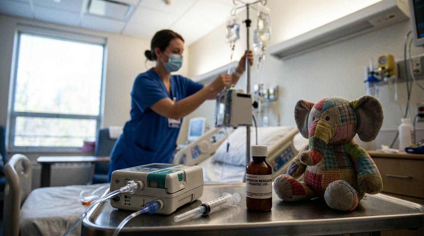
Infants with Diamond–Blackfan Anemia Syndrome and other congenital marrow failure disorders often require chronic red blood cell transfusions, a life-sustaining therapy that inevitably leads to secondary iron overload [1, 2]. Because the body lacks a physiological mechanism for excreting excess iron, the metal accumulates in vital organs, particularly the liver and heart, where it generates reactive oxygen species that cause progressive cellular damage [3, 4]. While chelation protocols are well-established for older children, the optimal timing for intervention in toddlers remains a clinical challenge, caught between the risks of early-onset organ damage and concerns about drug safety in very young patients [5, 6, 7, 8]. A recent multicenter study from France and Italy now provides critical real-world data on the feasibility, safety, and effectiveness of initiating chelation therapy in transfusion-dependent patients under three years of age.
Defining the Early Intervention Cohort
To evaluate iron removal in the youngest patients, investigators conducted a retrospective analysis using data from the French and Italian national registries for Diamond–Blackfan Anemia Syndrome. This approach allowed for the longitudinal tracking of patients with this rare congenital disorder across multiple centers. The study screened 167 transfused patients and identified a specific subgroup of 64 children, representing 38% of the total cohort, who had initiated chelation therapy before their third birthday. The median age for starting treatment in this early intervention group was 18 months, a critical developmental window where iron accumulation poses a significant systemic risk but for which clinical evidence has been historically sparse. This cohort provides a focused look at the outcomes of early chelation in a clinically vulnerable population.
Clinical Thresholds and Pharmacological Choices
The study identified two primary indications that prompted clinicians to begin chelation therapy in these young children: a serum ferritin level of at least 500 ng/mL and a history of more than 10 red blood cell transfusions. In practice, the degree of iron accumulation at the start of treatment was often substantial, with a median serum ferritin of 1340 ng/mL. This demonstrates that many toddlers had already developed a significant systemic iron burden by the time intervention was deemed necessary. Regarding the choice of chelating agents, the oral medication deferasirox was the most frequently used, prescribed for 63% of patients, a preference likely reflecting its ease of administration in a pediatric population. The parenteral agent deferoxamine was the second most common choice, utilized in 35% of patients. These real-world data on treatment triggers and drug selection offer a practical framework for managing iron overload in this age group.
Longitudinal Reduction in Systemic Iron Burden
Longitudinal analysis of the cohort confirmed that early intervention effectively controls systemic iron accumulation over time. The data showed that chelation was associated with a significant reduction in serum ferritin levels, declining at a rate of −11% per year (p < 0.001). This consistent annual decrease indicates that therapy initiated at a median age of 18 months can successfully counteract the iron load from ongoing transfusions, preventing its progression toward dangerously high levels. The sustained efficacy of this strategy was evident in follow-up data collected when the children reached 5 to 6 years of age. Among the 28 patients with available data at that time point, 43% had achieved ferritin levels below 500 ng/mL, indicating excellent control of iron stores. Critically, none of these patients had ferritin levels exceeding 2000 ng/mL, a key clinical objective, as concentrations above this threshold are strongly associated with an increased risk of cardiac and endocrine complications.
Organ Protection and Broader Clinical Utility
While serum ferritin is a useful biomarker, direct organ assessment is crucial for evaluating parenchymal damage. The investigators assessed liver iron concentration in 31 of the 64 patients (48%) at a median age of 3.2 years, revealing that 45% already had severe hepatic iron overload at their first evaluation. This finding underscores the rapidity with which iron can accumulate in the tissues of transfused infants. Despite this significant hepatic burden, the early initiation of chelation appeared to be highly effective in protecting the heart. Among 22 patients who underwent cardiac magnetic resonance imaging, no myocardial iron overload was detected. This suggests that starting chelation before age three provides a critical window to prevent cardiac iron deposition, the leading cause of mortality in chronically transfused patients. These real-world data support the feasibility and effectiveness of early chelation in this population and may help inform the management of iron overload in infants with other transfusion-dependent anemias, such as beta-thalassemia. The findings provide a strong rationale for developing age-specific chelation strategies to prevent irreversible organ damage.
References
1. Gloude NJ, Dickerson KE, Nakano TA. Congenital and Acquired Pediatric Bone Marrow Failure. Pediatrics in Review. 2025. doi:10.1542/pir.2024-006422
2. Liu Y, Karlsson S. Perspectives of current understanding and therapeutics of Diamond-Blackfan anemia. Leukemia. 2023. doi:10.1038/s41375-023-02082-w
3. Cheng HM, Holowka S, Moineddin R, Odame I. Liver iron overload assessment by T magnetic resonance imaging in pediatric patients: An accuracy and reproducibility study. American Journal of Hematology. 2012. doi:10.1002/ajh.23114
4. Duca L, Pierro ED, Scaramellini N, Granata F, Graziadei G. The Relationship Between Non-Transferrin-Bound Iron (NTBI), Labile Plasma Iron (LPI), and Iron Toxicity. International Journal of Molecular Sciences. 2025. doi:10.3390/ijms26136433
5. Patsourakos D, Aggeli C, Dimitroglou Y, et al. Speckle tracking echocardiography and β-thalassemia major. A systematic review. Annals of Hematology. 2023. doi:10.1007/s00277-023-05380-6
6. Kwiatkowski JL, Hamdy M, El‐Beshlawy A, et al. Deferiprone vs deferoxamine for transfusional iron overload in SCD and other anemias: a randomized, open-label noninferiority study. Blood Advances. 2021. doi:10.1182/bloodadvances.2021004938
7. Kotam GP, Morrison DB, Karikari JN, et al. Impact of sickle cell anaemia on reproductive health: a narrative review. Reproductive Health. 2025. doi:10.1186/s12978-025-02169-w
8. Hoffbrand AV, Taher A, Cappellini MD. How I treat transfusional iron overload. Blood. 2012. doi:10.1182/blood-2012-05-370098