Clinical Guideline Briefing
ERC ESICM Post-Resuscitation Care

ERC and ESICM launch 2025 guideline for Post-Resuscitation Care

New guidelines emphasize comprehensive diagnostic imaging, refined temperature control, and a multimodal approach to neurological prognostication after cardiac arrest.

ERC and ESICM launch 2025 guideline for Post-Resuscitation Care
Headline change
The guideline significantly updates the diagnostic approach for cardiac arrest etiology, prioritizing whole-body CT scans in specific non-ST-elevation scenarios.
Bedside action
For comatose post-ROSC patients without ST-elevation, consider immediate whole-body CT to identify treatable non-coronary causes before proceeding with coronary angiography.

The European Resuscitation Council (ERC) and the European Society of Intensive Care Medicine (ESICM) have released their 2025 guidelines for post-resuscitation care, building on the latest International Consensus on Cardiopulmonary Resuscitation Science. This update offers critical refinements across diagnosis, management, and prognostication, aiming to optimize outcomes for cardiac arrest survivors. Clinicians will find actionable guidance on navigating complex post-arrest scenarios, from immediate imaging strategies to long-term rehabilitation planning.

Versus the Previous Version

The 2025 ERC/ESICM guidelines for post-resuscitation care introduce several key updates, reflecting new evidence and a refined approach to patient management compared to the 2021 version. These changes impact initial diagnostic pathways, temperature management terminology, and long-term care strategies.

Cause of Cardiac Arrest Diagnosis
Previous version
Suggested coronary angiography first in patients with myocardial ischemia. CT brain and chest scan were considered if coronary angiography did not find causative lesions.
This version
Coronary angiography remains first if ST-elevation is present; otherwise, whole-body CT scan (including head, neck, chest, abdomen, pelvis, and CT pulmonary angiography) takes priority.
Clinical impact Clinicians should now consider a comprehensive whole-body CT scan earlier for non-ST-elevation patients to identify non-coronary causes, potentially altering the diagnostic sequence.
Oxygenation Management
Previous version
Recommendation to start with 100% oxygen immediately after ROSC, then titrate to 94–98% SpO2 or PaO2 10–13 kPa (75–100 mmHg).
This version
Maintains recommendation and adds explicit guidance highlighting inaccuracies in pulse oximetry in patients with darker skin tones.
Clinical impact While oxygen targets remain similar, be aware of potential pulse oximetry inaccuracies in patients with darker skin tones and low-flow states, requiring careful clinical correlation.
Coronary Reperfusion Strategy
Previous version
Suggested delaying cardiac catheterisation if clinical context does not clearly indicate a high likelihood of acute coronary occlusion in OHCA patients without ST-elevation.
This version
Immediate coronary angiography strongly considered in OHCA without ST-elevation if high likelihood of coronary occlusion.
Clinical impact The threshold for immediate coronary angiography in OHCA patients without ST-elevation is lowered, requiring careful assessment of clinical likelihood of coronary occlusion.
Temperature Management
Previous version
Recommended targeted temperature management at 32–36°C for at least 24 h and fever avoidance (>37.7°C) for at least 72 h post-ROSC.
This version
Preferred terminology is temperature control. Recommends actively preventing fever 37.5°C for at least 72 h post-ROSC.
Clinical impact The terminology shifts from 'targeted temperature management' to 'temperature control,' with a specific focus on active fever prevention (target <37.5°C) for a longer duration (at least 72 hours).
Neurological Prognostication
Previous version
Emphasised multimodal neurological assessment at 72 h.
This version
Maintains recommendation with specified indicators of favourable neurological outcome and suggested timing for brain CT and SSEP recording added to the algorithm.
Clinical impact The prognostication algorithm is enhanced with explicit indicators for favorable neurological outcomes and more precise timing for diagnostic tests like brain CT and SSEP.
Rehabilitation and Follow-up
Previous version
Recommended functional assessment before discharge and follow-up within 3 months post discharge including screening of cognitive, emotional problems and fatigue. Brain injury and cardiac rehabilitation when indicated.
This version
Maintains recommendations and adds structured guidance on rehabilitation in the ICU including early mobilisation, delirium management, ICU diaries, and to address physical limitations during follow-up. Stronger focus on the involvement of co-survivors.
Clinical impact Rehabilitation guidance is expanded to include specific ICU interventions like early mobilization and delirium management, with increased emphasis on supporting co-survivors.
Organ Donation
Previous version
Recommended considering organ donation post-resuscitation.
This version
Maintains recommendation and adds recommendations for cardiac arrest registries to report organ donation activities.
Clinical impact Beyond considering organ donation, there's a new emphasis on reporting organ donation activities in cardiac arrest registries to improve data collection.
Unexplained Cardiac Arrest
Previous version
Not included.
This version
New recommendations for comprehensive diagnostic work-up (including genetic testing, cardiac MRI, sodium channel blocker tests, exercise testing) and emphasises long-term follow-up.
Clinical impact A new section provides detailed guidance on investigating unexplained cardiac arrest, requiring a comprehensive diagnostic workup and long-term follow-up for these patients.

Practice Notes

These practical considerations highlight critical aspects of post-resuscitation care that demand careful attention in daily practice.

Caution
Be aware of pulse oximetry inaccuracies in patients with darker skin tones or low-flow states.
Pulse oximetry can overestimate true oxygen saturation in these populations, potentially masking hypoxemia. Clinical judgment and arterial blood gas measurements are crucial for accurate oxygenation assessment.
Implementation
Prioritize immediate whole-body CT for comatose OHCA patients without ST-elevation if non-coronary causes are suspected.
This updated diagnostic pathway aims to rapidly identify treatable causes like pulmonary embolism or hemorrhage before considering coronary angiography, optimizing early intervention.
Monitoring
Actively prevent fever by targeting a temperature <37.5°C for at least 72 hours in comatose post-ROSC patients.
Fever prevention is now explicitly recommended for a longer duration, emphasizing continuous temperature monitoring and control to mitigate secondary brain injury.
Shared decision point
Separate discussions about withdrawal of life-sustaining treatment (WLST) from neurological prognostication.
WLST decisions should encompass broader factors like age, comorbidities, and patient preferences, not solely brain injury prognosis, to avoid premature withdrawal.
Do not miss
Implement early mobilization, delirium management, and ICU diaries during hospitalization for cardiac arrest survivors.
These interventions are crucial for improving long-term functional outcomes and addressing physical and cognitive impairments post-ICU stay.
Special population
Consider mechanical circulatory support for persisting cardiogenic shock in select patients, such as those with STEMI and short cardiac arrest duration.
While not routinely recommended, MCS can be life-saving in specific subgroups with refractory shock, especially when brain injury risk is low.

Key Recommendations

The following key recommendations from the ERC/ESICM 2025 guidelines provide actionable guidance for managing adult patients after cardiac arrest. They cover critical decisions from initial diagnosis and physiological support to neurological prognostication and long-term recovery, aiming to improve patient outcomes in daily clinical practice.

Key recommendations14 highlighted
01

In adult patients with ROSC after cardiac arrest of suspected cardiac origin with persistent ST-elevation on the ECG., perform emergent cardiac catheterisation laboratory evaluation (and primary percutaneous coronary intervention (PPCI) if required).

Line / StrengthStrong
Level of EvidenceLow-quality evidence (from 2021 guideline, implied unchanged)
EvidenceThis recommendation is based on strong evidence for the benefit of early PCI for STEMI without cardiac arrest, and observational studies showing benefit for emergent PCI in post-ROSC patients with ST-elevation. An ILCOR evidence update in 2025 identified no additional studies involving participants with ST-elevation to change this. The 2023 ESC guidelines also support this approach.
For practiceExpedite patients with ST-elevation post-ROSC to the cath lab for immediate evaluation and intervention.
CaveatST-elevation on post-ROSC ECG can be transient; repeat ECG on hospital arrival is important.
02

In adult patients with ROSC after out-of-hospital cardiac arrest (OHCA) without ST-elevation on the ECG, where clinical context suggests a high likelihood of acute coronary occlusion., strongly consider immediate coronary angiography.

Line / StrengthStrongly considered
Level of EvidenceNot specified, but implies expert consensus based on clinical context.
EvidenceWhile large RCTs (COACT, TOMAHAWK) showed no benefit for immediate routine angiography in OHCA without ST-elevation, this recommendation applies to cases with a 'high likelihood of coronary occlusion' based on factors like hemodynamic/electrical instability, pre-arrest symptoms, and ECG patterns, aligning with a nuanced approach.
For practiceIndividualize the decision for immediate angiography in non-ST-elevation OHCA based on a comprehensive clinical assessment.
CaveatDelaying angiography may be reasonable if the clinical context does not strongly suggest acute coronary occlusion.
03

In patients with signs or symptoms pre-arrest suggesting a non-coronary cause (e.g., headache, seizures, neurological deficits, shortness of breath, documented hypoxemia, abdominal pain)., perform a dual phase whole body tomography (CT) scan (including head, neck, chest, abdomen, pelvis, and CT pulmonary angiography) before or after coronary angiography if indicated.

Line / StrengthPerform
Level of EvidenceExpert consensus (from 2021 guideline, implied unchanged)
EvidenceThis recommendation is based on expert consensus and observational studies showing that early CT imaging can identify extracardiac causes in a substantial proportion of patients, sometimes making specific therapeutic intervention possible. Systematic whole-body CT has shown to increase diagnostic yield and shorten time to diagnosis without increased complications.
For practiceIntegrate comprehensive CT imaging early in the diagnostic workup for patients with suspected non-cardiac causes of arrest.
CaveatThe timing relative to coronary angiography depends on the clinical indication and suspicion of cardiac versus non-cardiac etiology.
04

In patients who remain comatose following ROSC, or who have another clinical indication for sedation and mechanical ventilation., have their trachea intubated if this has not been done already during CPR.

Line / StrengthShould
Level of EvidenceExpert consensus (from 2021 guideline, implied unchanged)
EvidenceTracheal intubation facilitates controlled oxygenation and ventilation, protects against aspiration, and allows for management of seizures, temperature, and brain injury in critically ill, comatose patients post-ROSC.
For practiceEnsure timely and skilled tracheal intubation for comatose or mechanically ventilated post-ROSC patients.
CaveatIntubation should be performed by experienced operators to ensure high success rates and confirmed with waveform capnography.
05

In adults with ROSC after cardiac arrest in any setting, once SpO2 can be measured reliably or arterial blood gas values are obtained., titrate the inspired oxygen to achieve an arterial oxygen saturation of 94–98% or arterial partial pressure of oxygen (PaO2) of 10–13 kPa (75–100 mmHg).

Line / StrengthSuggest targeting
Level of EvidenceGood practice statement
EvidenceThis guidance is based on aiming for 'normal' oxygenation values (normoxia), with evidence of harm from hypoxemia and potential harm from hyperoxemia. Randomized trials have not identified an optimal target but support normoxemia as safe.
For practiceActively manage inspired oxygen to maintain normoxemia, using ABGs for precise titration and considering pulse oximetry limitations.
CaveatBe aware that pulse oximetry can overestimate true oxygen saturation in people with darker skin tones and in low-flow states.
06

In mechanically ventilated patients with ROSC after cardiac arrest., target normocapnia – a partial pressure of carbon dioxide of 4.7–6.0 kPa (or approximately 35–45 mmHg).

Line / StrengthTarget
Level of EvidenceExpert consensus (from 2021 guideline, implied unchanged)
EvidenceWhile evidence for a specific PaCO2 target is inconsistent, normocapnia is generally recommended. Hypercapnia increases cerebral blood flow and intracranial pressure, while hypocapnia can cause cerebral vasoconstriction and ischemia. Lung protective ventilation strategies are also recommended.
For practiceMaintain PaCO2 within the normocapnic range, adjusting ventilation settings and monitoring closely, especially in hypothermic patients.
CaveatMonitor PaCO2 frequently in hypothermic patients as hypocapnia may occur; use consistent temperature or non-temperature corrected blood gas values.
07

In all patients after cardiac arrest., avoid hypotension and target a mean arterial pressure (MAP) >60–65 mmHg.

Line / StrengthAvoid and target
Level of EvidenceWeak recommendation, based on weak evidence (from 2021 guideline, implied unchanged)
EvidenceA systematic review including over 1000 patients found a higher MAP target was not associated with higher survival or better functional outcomes but also no significant risks. Observational studies consistently link MAP values below 60-65 mmHg with higher mortality. ILCOR made a weak recommendation to target MAP >60-65 mmHg.
For practiceMaintain MAP above 60-65 mmHg, individualizing targets based on patient specifics and signs of perfusion.
CaveatA higher MAP target might be appropriate in selected patients (e.g., chronic hypertension, persistent peripheral hypoperfusion).
08

In patients with arrhythmia immediately after ROSC., follow the ERC Guidelines 2025 Adult Advanced Life Support for peri-arrest arrhythmia.

Line / StrengthFollow
Level of EvidenceExpert consensus
EvidenceThis recommendation directs clinicians to the comprehensive ERC ALS guidelines for immediate management of arrhythmias, acknowledging the complexity and specific protocols required for these events.
For practiceRefer to ALS guidelines for acute arrhythmia management and actively investigate and treat precipitating factors.
CaveatTreat any potential underlying causes, such as coronary occlusion or electrolyte disorders.
09

In patients who remain comatose after ROSC from cardiac arrest., actively prevent fever by targeting a temperature <37.5 °C.

Line / StrengthSuggest actively preventing
Level of EvidenceWeak recommendation, low-certainty evidence
EvidenceILCOR's systematic reviews found no difference between hypothermic and normothermic temperature control, but suggest actively preventing fever. Observational studies link fever to worse outcomes. The recommendation is a weak one due to low certainty evidence.
For practiceImplement strategies to maintain normothermia and prevent fever, such as surface or endovascular cooling devices with feedback systems.
CaveatComatose patients with mild hypothermia (32–36°C) after ROSC should not be actively warmed to achieve normothermia.
10

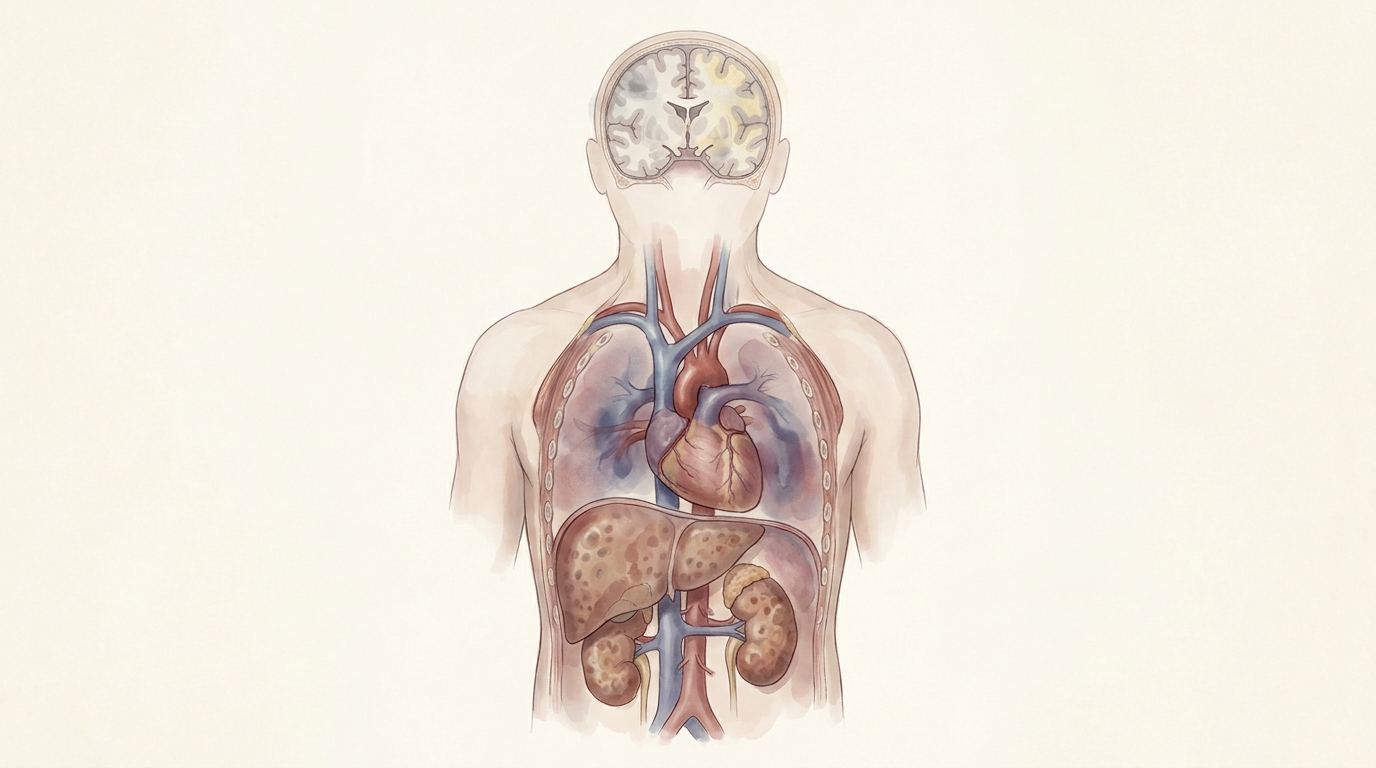
In comatose patients after resuscitation from cardiac arrest., perform neurological prognostication using clinical examination, electrophysiology, biomarkers, and imaging.

Line / StrengthShould be performed
Level of EvidenceExpert consensus (from 2021 guideline, implied unchanged)
EvidenceNo single predictor is 100% accurate, necessitating a multimodal approach to inform relatives and guide treatment decisions, aiming for high specificity to avoid falsely pessimistic predictions.
For practiceIntegrate multiple diagnostic modalities for a comprehensive and accurate neurological prognosis.
CaveatExclude potential residual effects of sedatives and other drugs that may confound test results.
11

In unconscious patients at 72 hours from ROSC, in the absence of confounders, where poor outcome is likely., identify poor outcome if two or more of the following predictors are present: no pupillary and corneal reflexes at 72h, bilaterally absent N20 SSEP wave at 24h, highly malignant EEG at >24h, NSE >60 mg/L at 48h and/or 72h, status myoclonus 72h, or diffuse and extensive anoxic injury on brain CT/MRI.

Line / StrengthVery likely
Level of EvidenceStrong recommendation, very low-certainty evidence (from 2021 guideline, implied unchanged)
EvidenceThis multimodal prognostication algorithm, validated in multicenter studies, predicts poor outcome with a 0% false positive rate when two or more concordant signs are present. The specificity of individual tests varies, but their combination enhances reliability.
For practiceApply this multimodal algorithm systematically at 72 hours post-ROSC to guide discussions and decisions regarding prognosis.
CaveatMost signs can be recorded before 72h, but results are evaluated at the time of clinical prognostic assessment.
12

In patients who are comatose after cardiac arrest., use electroencephalography (EEG) to diagnose electrographic seizures in patients with clinical convulsions and to monitor treatment effects.

Line / StrengthRecommend using
Level of EvidenceNot specified (from 2021 guideline, implied unchanged)
EvidenceEEG is a widely used and studied method for assessing brain function and prognosis, and is essential for diagnosing and treating seizures, which are reported in 20-30% of cardiac arrest patients.
For practiceIntegrate EEG monitoring for seizure detection and treatment response, especially in comatose patients.
CaveatContinuous EEG monitoring is labor-intensive and may add significant cost.
13

In adult patients with non-traumatic OHCA., consider for transport to a cardiac arrest centre for post-resuscitation care, whenever possible, according to local protocols.

Line / StrengthShould be considered
Level of EvidenceWeak recommendation, low-certainty evidence
EvidenceThis recommendation is based on low-certainty evidence from a systematic review, which included one RCT and 15 observational studies. While most studies reported a survival benefit with care at a cardiac arrest center, heterogeneity and risk of bias were significant.
For practiceFacilitate transfer of eligible OHCA patients to specialized cardiac arrest centers to optimize post-resuscitation care delivery.
CaveatThe optimal configuration of cardiac arrest centers varies, and local protocols should guide implementation.
14

In all patients who have restoration of circulation after CPR and who subsequently progress to death., be evaluated for organ donation.

Line / StrengthRecommend that
Level of EvidenceExpert consensus
EvidenceRecent CPR is not a barrier to organ donation, and a significant proportion of non-surviving patients represent potential organ donors. Organ donation policies and practices vary internationally, requiring adherence to local legal and ethical requirements.
For practiceIntegrate organ donation evaluation into end-of-life care pathways for all eligible post-ROSC patients.
CaveatAll decisions concerning organ donation must follow local legal and ethical requirements.

SOURCE GUIDELINE

European Resuscitation Council and European Society of Intensive Care Medicine Guidelines 2025 Post-Resuscitation Care

ERC/ESICM

Year2025
TypeFull Guideline
JournalResuscitation
DOI10.1016/j.resuscitation.2025.110809